Usually searching “How the eyes work” leads you only to phototransduction which only concerns rods, cones and retinal photosensitive ganglion cells. But this is not everything from A to Z about how the eye works, is it? The eye is a whole organ with many wonders, whether it’s a structure, a muscle/fiber or a nerve/neuron. To clarify, when each tiny part has a significant role, then all parts go under the same title “How the eyes work”. So its not just how the eye converts the light into electrical signals, it also includes how the eye moves. Therefore, below we’ll discuss the literal concept of the eye, from how it moves to how we see.
How the eyes move:

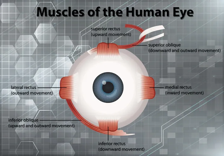
Six extraocular muscles attaches to the eye in the eye socket or orbit. In fact, these muscles move the eye up and down, right and left, and rotate it.
- Superior oblique: Provides an angular attachment to the posterior side of the eye. To illustrate, it’s responsible for down-and-out eye movement and intortion (incycloduction) of the eye. The superior oblique originates within the periosteum of the sphenoid and passes along the medial border of the orbital roof before reaching the trochlea. A side note, the trochlea is a fibrous, cartilaginous pulley which resides on the trochlear fovea of the frontal bone. Once at the trochlea, its tendon turns postero-laterally and crosses the eyeball until it reaches its insertion point in the outer posterior quadrant of the eye. The cranial nerve IV or trochlear nerve supports this muscle.
- Inferior oblique: It’s the shortest muscle relative to other extraocular muscles. External rotation is the primary purpose of the inferior oblique muscle, while elevation and abduction are its secondary and tertiary functions. Located just lateral to the nasolacrimal groove, the inferior oblique arises from the orbital floor, and it’s innervated by the cranial nerve III known as the oculomotor.
- Superior rectus: Attached vertically to the eyeball at the top. Indeed, this muscle is responsible for eye elevation primarily, and it may be involved in adduction and intortion due to it making 23 degrees with the visual axis. The superior rectus extends superiorly and anteriorly over the eyeball, and originates from the annulus of Zinn. Finally, it’s innervated by the cranial nerve III (oculomotor).
- Inferior rectus: Similarly, the inferior rectus also originates from the annulus of Zinn. However, the primary role of this muscle is eye depression. Also, because it extends laterally and anteriorly over the eyeball and makes 23 degrees with the visual axis, it does abduction and extorsion (excyloduction) as well. Moreover, the oculomotor is the nerve within this muscle.
- Medial rectus: In fact, Its primary role is adduction. It originates from the tendinous ring. Besides, the medial rectus is innervated by the oculomotor never.
- Lateral rectus: With abduction being its role in eye movement, the lateral rectus along with the medial rectus allows the lateral movement or side to side movement of the eye. Furthermore, it originates from the annulus of Zinn and it’s innervated by the abducens nerve (CN IV).
How vision works (Visual phototransduction):
Before we go into details, its important to mention two neurons (photoreceptors) involved in visual phototransduction.
Rod cells:
With an average of 92 million rod cells in the human retina, these cells are light sensitive. To clarify, just one photon is enough to stimulate rod cells, making vision possible in the nighttime or dusk when light is very little. Moreover, as their name indicates, rode cells are rod like elongated thin cells made of two segments. Rod cells are stacked as discs, each disc includes rhodopsin protein (visual purple).
Cone cells:
These are not as numerous as rod cells; An average of 4.6 million cells are inside the human retina, and they work in bright light allowing us to see colors. There are 3 classes on cones, each class has a different iodopsin (visual pigment) type. The classes are:
- Short-wavelength cones (For blue color observation)
- Medium-wavelength cones (For green light vision)
- Long-wavelength cones (For red light observation)
Phototransduction:
To begin with, the retinal surface is parallel to the stacked membranous discs of rods and cones, so light can reach these regions easily. Both rhodopsin and iodopsin proteins resides in the membranes in high concentration. The photoreceptor cells of the retina contains a transmembrane protein known as the opsin, in addition to a bound light-sensitive chromophore molecule. The chromophore in rhodopsin is known as retinal which is a derivative of vitamin A.
Phototransduction is a process in which changes happen in the cells when light strikes and activates the chromophore, a process common to rods and cones. In darkness, rhodopsin plays no role and cation channels are open. Darkness inhibits rhodopsin activity and opens cation channels in the cell membrane. The cell depolarizes and continuously releases neurotransmitters at the synapse with the bipolar neurons. Upon absorption of a photon of light, retinal isomerizes from 11-cis- to all-trans-retinal within one picosecond; this causes configuration changes for the opsin that ultimately activate transducin, an adjacent membrane-associated heterotrimeric G protein (opsin is couples with this protein).
As a result, transducin activity induces hyperpolarization that reduces the release of neurotransmitters at the synaptic junction of neurons, and this in turn depolarizes sets of bipolar neurons. Consequently, the optic nerve ganglion cells receives action potentials from these depolarized cells. This process of confirmation change in retinal induced by light also leads to the dissociation of the chromophore from the opsin, known as bleaching. In the adjacent pigmented epithelial cell, all-trans-retinal is converted back into 11-cis-retinal, then transported back into the photoreceptor, where it is reabsorbed. In this cycle, the retina regenerates and the rhodopsins recover from the bleaching, which is part of the slow adaptation of the eyes to a change in light level. To sum it up, light signals changes to electrical signal that passes through the optic nerve into the brain which shows us images.
References:
-Abdelhady A, Patel BC, Aslam S, et al. Anatomy, Head and Neck, Eye Superior Oblique Muscle. [Updated 2021 Aug 11]. In: StatPearls [Internet]. Treasure Island (FL): StatPearls Publishing; 2022 Jan-.
-Gupta N, Patel BC. Anatomy, Head and Neck, Eye Inferior Oblique Muscles. [Updated 2021 Jul 31]. In: StatPearls [Internet]. Treasure Island (FL): StatPearls Publishing; 2022 Jan-.
-Shumway CL, Motlagh M, Wade M. Anatomy, Head and Neck, Eye Superior Rectus Muscle. [Updated 2021 Jul 26]. In: StatPearls [Internet]. Treasure Island (FL): StatPearls Publishing; 2022 Jan-.
-Shumway CL, Motlagh M, Wade M. Anatomy, Head and Neck, Eye Inferior Rectus Muscle. [Updated 2021 Jul 26]. In: StatPearls [Internet]. Treasure Island (FL): StatPearls Publishing; 2022 Jan-.
-Shumway CL, Motlagh M, Wade M. Anatomy, Head and Neck, Eye Medial Rectus Muscles. [Updated 2021 Sep 17]. In: StatPearls [Internet]. Treasure Island (FL): StatPearls Publishing; 2022 Jan-.
-Mescher, A. L., Mescher, A. L., & Junqueira, L. C. U. (2016). Junqueira's basic histology: Text and atlas (Fourteenth edition.). New York: McGraw-Hill Education.


